Directly atomic observation of zeolites and zeotypes
- Abstract number
- 53
- Event
- European Microscopy Congress 2020 Invited Speakers
- DOI
- 10.22443/rms.emc2020.53
- Corresponding Email
- [email protected]
- Session
- PSA.8 - Microscopy in industrial applications
- Authors
- Dr Alvaro Mayoral (1)
- Affiliations
-
1. Center for High-resolution Electron Microscopy (CћEM), School of Physical Science and Technology, ShanghaiTech University
- Keywords
Beam Damage, Electron Microscopy, MOFs, Zeolite
- Abstract text
Ordered nanoporous materials, where zeolites and derivate are the most representative ones, are heavily used in industry mainly as ion exchangers and as heterogeneous catalysts. Applications that are continuously being expanded owed to their particular structural features such as well‐defined porous network, thermal stability, acidity or guest-specimen confinement.
Zeolitic physicochemical properties are directly related to the structure, which can be described as tetrahedra TO4 sharing oxygen bridges (T = Si, Al), that can generally be described as Mm+x/m[Si1-xAlxO2], where M is an exchangeable counter cation with valence m+ to balance the negative framework charge, and the range of x is equal to or less than 0.5 and can be as low as 0.0 for pure silica polymorphs. In order to understand these structural related properties, a deep atomic characterization is highly required. Electron microscopy (EM) could give response to what nowadays is necessary in order to produce new functional materials with tuned properties.
Traditionally, high-resolution transmission electron microscopy (HRTEM) has been limited to the observation of pores and few cases to identify some of the member rings that form the zeolites in terms of size, shape and periodicity. These restrictions have been attributed to the spatial resolution of the electron microscopes and to the very high insatiability of these materials respect to the electron beam resulting in a complete structural disintegration within just seconds of observation[1]. With the implementation of the aberration correctors in both scanning/transmission electron microscopes, (S)TEM[2], sub-Ångstrom lateral atomic resolution has been achieved for e-beam stable inorganic materials. Despite the difficulties that can be encountered for the analysis of zeolites and zeotypes, the motivation of using electron microscopy, especially in STEM configuration, is related to their widespread use as supports for metallic species either in their pores or on their external surfaces. However, location of the clusters and/or atoms have not been so evident since T-atoms of the framework are not usually resolved.
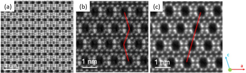
A good example of the minimum requirements for the characterization and developments of the next generation of functional porous materials, concerns to the analysis of all T-atoms (Al and P) of an e-beam sensitive AlPO4 (STA- 20)[3]. By a multitechnique approximation, the structure was solved and the complete framework was directly imaged by Cs-corrected STEM using an annular dark field, ADF, detector (figure 1(a)). Such a level of information is also required to evaluate ion-exchanged materials or highly defective ones. This is the case for example of the new zeolite, HPM-8, a germanosilicate from the Beta family formed by the polymorphs D and E[4], see figure 1(b) and 1(c).
Figure
1. Cs-corrected STEM-ADF observations of (a) STA-20 silicoaluminophosphate. (b) and (c) two polymorphs found in HPM-8 material observed along the same zone axis.
Alongside these structural analyses on new materials, the current challenges in the analysis of beam sensitive porous structures rely on the observation of oxygen bridges, which has been achieved in various structures with special emphasis on one of the most e-beam sensitive and heavily used zeolites, Na-LTA. Furthermore, together with the observation of ion-exchanged zeolites the results on the observation of single transition metal (Fe) heteroatoms within the MFI framework will be also presented and discussed[5].
In summary, electron microscopy can give response to many questions related to ordered porous materials used in industry. It is expected that the newer technologies that are currently being developed such as dedicated annular bright field (ABF) detectors, phase image retrieving in STEM mode or direct detection electron counting cameras can provide further insights on zeolitic science[6].
- References
[1] I. Díaz and A. Mayoral, Micron, 42 (2011) 512–527.
[2] O. Krivanek, N. Dellby, A. J. Spence, R. A. Camps and L. M. Brown, In Proc. EMAG 1997, Cambridge, UK (1997) 35–49.
[3] A. Turrina, R. Garcia, A. E. Watts, H. F. Greer, J. Bradley, W. Zhou, P. A. Cox, M. D. Shannon, A. Mayoral, J. L. Casci and P. A. Wright, Chem. Mater. 29 (2017) 2180–2190.
[4] P. Lu, A. Mayoral, L. Gómez-Hortigüela, Y. Zhang and M. A. Camblor, Chem. Mater. 15 (2019) 5484-5493.
[5] A. Mayoral, Q. Zhang, Y. Zhou, P. Chen, Y. Ma, T. Monji, P. Losch, W. Schmidt, F. Schüth, H. Hirao, J. Yu and O. Terasaki, submitted.
[6] The authors gratefully acknowledge funding from The Centre for High-resolution Electron Microscopy (CħEM), supported by SPST of ShanghaiTech University under contract No. EM02161943; to the Natural National Science Foundation of China NFSC-21850410448, NSFC- 21835002.