Label-free Multimodal Nonlinear Microscopy to Assess Stem Cell Differentiation in 3D Nichoids
- Abstract number
- 1097
- Event
- European Microscopy Congress 2020
- DOI
- 10.22443/rms.emc2020.1097
- Corresponding Email
- [email protected]
- Session
- LSA.1 - Label-free life science imaging
- Authors
- Ms Valentina Parodi (1), Ms Benedetta Talone (3), Ms Arianna Bresci (2), Dr Emanuela Jacchetti (2), Dr Tommaso Zandrini (5), Prof Roberto Osellame (6), Prof Giulio Cerullo (4), Prof Dario Polli (4), Prof. Manuela Raimondi (2)
- Affiliations
-
1. Department of Chemistry, Materials and Chemical Engineering, “G. Natta”, Politecnico di Milano 20133, Italy
2. Department of Chemistry, Materials and Chemical Engineering, “G. Natta”, Politecnico di Milano 20133, Italy;
3. Department of Physics, Politecnico di Milano, 20133 Milano, Italy
4. Department of Physics, Politecnico di Milano, 20133 Milano, Italy;
5. Institute of Material Science and Technology, Technische Universität Wien, 1040 Vienna, Austria;
6. Istituto di Fotonica e Nanotecnologie (IFN)-CNR, 20133 Milano, Italy
- Keywords
CARS, Multimodal Microscopy, SHG, Stem Cells
- Abstract text
Summary
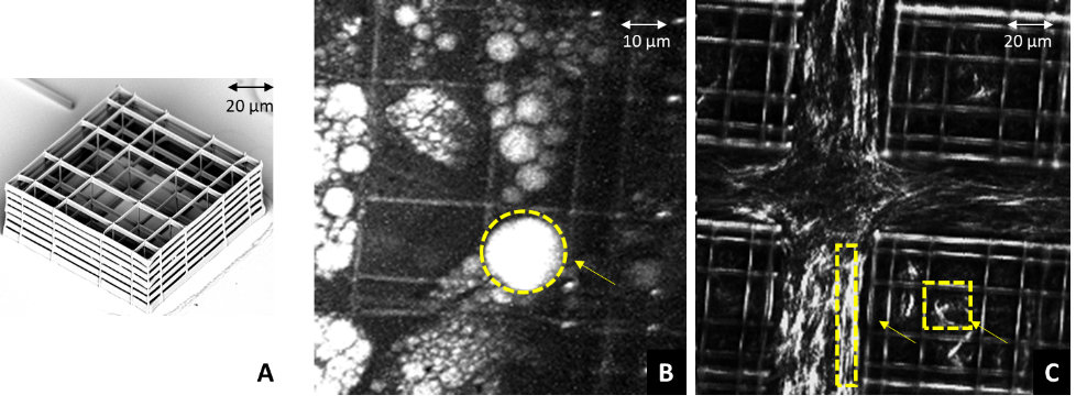
We employed a novel multimodal nonlinear microscope [1] to characterize the 3D Nichoid platform [2] (fig.1A). Our system provides two-photon excitation fluorescence (TPEF), second harmonic generation microscopy (SHG) and coherent anti-Stokes Raman scattering (CARS). Here, we applied multimodal microscopy to investigate stem cells differentiation towards adipogenic and chondrogenic phenotype inside Nichoids.
Introduction
A recent challenge in bioimaging field is the possibility to optically monitor in real-time vital, thick and complex tissues in their native three-dimensional state. Among different diagnostic techniques, we found out nonlinear optical microscopy as the best non-invasive method for assessing cellular physiology.
Materials and Methods
CARS in the CH-stretch region (2500-3200cm-1) was obtained from the overlap of pump (780nm) and Stokes (940-1200nm) pulses. TPEF/SHG signals were obtained from a 780nm and 100fs pulsed laser.
Results and Discussion
Figure 1B shows CARS signal at 2845cm-1 for adipogenesis inside the Nichoid, while in figure 1C collagen fibrils outside and inside the Nichoid.
Conclusion
Our study underlined the potentiality of multimodal microscopy to study physio pathological processes in vital and thick biological samples without staining, thus representing a turning point towards clinical studies.
Figure 1:A) SEM image of the elementary unit of 3D Nichoid scaffold. B) CARS image at 2845 cm-1 of lipid vesicles produced in adipogenesis and C) SHG image of collagen fibrils after chondrogenesis inside Nichoids.
- References
1. Crisafi F et al. Spectrochim Acta A Mol Biomol Spectrosc 2017;188:135-140
2. Nava, M. M. et al. Stem Cell Res. Ther. 7, 1–12 (2016).

